Services
Services we deliver
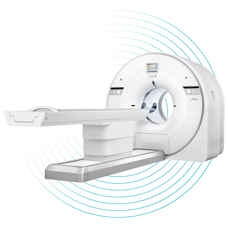
Mobile Imaging is bringing comprehensive care close to home by providing advanced diagnostics through a mobile PET-CT service for Aotearoa New Zealand.
There are two broad types of scans that can be completed on the mobile unit: standard CT and PET-CT scans.

CT Scan
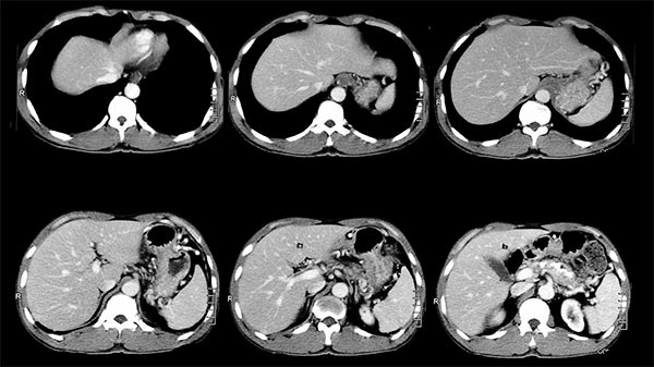
The CT (Computed Tomography) scanner uses computer processing and X-ray to produce detailed cross-sectional images of the body, including three-dimensional pictures. Unlike standard X-rays, CT scan show bones, soft tissue anatomy, blood vessels and air – all in very fine detail. A radiologist interprets these images. CT imaging is a highly advanced form of X-ray beam that shows the anatomical detail inside the body and detects alterations of structure caused by disease.
PET-CT Scan
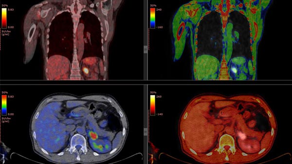
Positron Emission Tomography (PET) is an imaging procedure that uses small amounts of radioactive tracers to help diagnose, locate and assess a disease. It can be used to study specific areas or the whole body. The functional PET images are fused with high-definition anatomical CT images, after which the scan is called a PET-CT scan. On the mobile unit, two types of radioactive pharmaceuticals are used, including FDG for all body scanning and PSMA for prostate cancer imaging.
CT scan
What is a CT scan?
A CT scan, short for computerised tomography, is an imaging tool that helps doctors see detailed pictures of the inside of your body. Here’s how it works:
- X-Ray images from different angles: During a CT scan, you lie on a table that moves through a circular machine. This machine takes a series of X-ray images from different angles around your body.
- Cross-Sectional slices: These X-ray images are then processed by a computer to create cross-sectional slices. Each slice shows a different layer of your body.
- Detailed information: These slices reveal detailed information about your bones, blood vessels, and soft tissues. Much more than what regular X-rays can show!
- Diagnosis and treatment planning: Doctors use CT scans to diagnose diseases, check for injuries, and plan medical treatments. For example, they can determine whether a patient has a broken bone, a tumor, or other health issues.
Remember, a CT scan is a valuable tool that helps doctors take a closer look inside your body to keep you healthy!
CT SCAN
A CT scan is a medical test that uses X-rays to create detailed images of the inside of your body, helping doctors diagnose diseases and plan treatments.

What is a CT-Scan
A CT scan, or Computed Tomography scan, is a type of medical imaging that uses X-rays to create detailed pictures of the inside of your body.
Why are CT scans done?
Your doctor may ask for a CT scan to help:
- identify the cause of unexplained weight loss, pain or a lump that you or your doctor can feel
- identify muscle and bone problems, such as injuries or fractures
- monitor the progress of diseases, eg, heart disease or cancer
- identify issues with your blood vessels, gall bladder, liver or bladder
- identify the exact location of an infection or a blood clot
- guide procedures such as biopsies, surgery and radiation treatment
- detect internal bleeding or injuries.
Will I be exposed to radiation during a CT scan?
Yes, CT scans do use radiation, but the amount is low and is considered safe for most people. The benefits of getting accurate diagnostic information usually outweigh the potential risks.
What is the difference between a CT scan and an MRI?
Both CT scans and MRIs can create detailed images of the body, but they use different technologies. CT scans use X-rays, while MRIs use strong magnetic fields and radio waves. Your doctor will decide which is best for you based on your specific needs
How will I get my results?
A radiologist will look at your scan and will send the results to your doctor.
PET-CT scan using FDG
What is a PET scan?
A Positron Emission Tomography (PET) scan is a remarkable imaging test that provides insights into how your organs and tissues function. Unlike static images, PET scans capture dynamic processes within your body. Here’s how it works:
Patients receive a tiny amount of a radioactive substance (called a tracer) through an injection. This tracer is like a spotlight that highlights areas with active metabolism. Patients sit resting for a period of time to allow the tracer to move throughout their bodies. It is important to avoid strenuous movements during this time, as they can affect where the tracer accumulates in their bodies.
Once injected, the radiotracer mingles with your tissues. It emits positrons, which are positively charged particles. These positrons collide with electrons in your body, resulting in a ‘dance’ of energy conversion. When a positron meets an electron, they annihilate each other in a burst of energy. This energy is emitted as gamma rays. The PET scanner, a sophisticated device, detects these gamma rays. The scanner creates detailed images based on the distribution of gamma rays.
The Magic of PET-CT: Combining Forces
PET scans alone show metabolism but not precise anatomy, so that’s why our scanner has both a CT scanner and a PET scanner combined. The CT takes detailed X-ray images of your body’s structure. The PET takes information on metabolic function. Combining PET and CT reveals both metabolic function (like PET) and precise location (like CT).
FDG
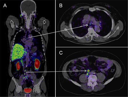
There is a wide array of different radioactive tracers used for different specialty purposes, but the mobile unit utilises FDG (18F-FDG) to investigate the whole body, specifically in the area of oncology (cancer). The role of this type of scan may be to stage and monitor the response to therapy for malignant disease.
FDG for all-body scans
FDG (fluorodeoxyglucose) is the most common radioactive glucose used to identify a wide range of cancers throughout the body during a PET-CT scan. The glucose very accurately detects areas of high metabolic activity so is a very widely used tracer on the mobile unit.

Why are PET-CT scans done?
A PET scan is done to help diagnose, stage or monitor a wide range of diseases. These include:
- Cancer staging or restaging – evaluating the extent of cancer spread, checking whether treatment is working or finding a recurrence.
- Cancer detection – in many different organs, eg, brain, breast, lung, prostate, skin, colorectal, lymph system.
- Heart disease – finding areas of abnormality, eg, sarcoidosis.
- Brain disorders – evaluation of brain tumours, Alzheimer’s disease and seizures.
What happens during the PET scan?
The procedure and the amount of time it takes is different depending on the reason for the scan and the part of the body being scanned. You’ll be given information about what to expect before you have your scan.
Generally, you will be given the radiotracer injection 45–60 minutes (called uptake time) before the scan and will have to rest quietly while the tracer circulates within your body.
After the appropriate amount of uptake time, you will lie on a padded table that slides into the short tunnel of the PET/CT scanner. It’s important that you keep it very still so the images are not blurry. The scanner is relatively quiet most of the time. You won’t feel any pain during the scan. Contrast for the CT scan may give you a momentary warm sensation or metallic taste in your mouth.
The entire procedure usually takes up to 2 hours.
What happens after the scan?
- The amount of radioactive tracer used during the PET scan is small and will be flushed out of your system in a few hours.
- Drinking plenty of fluids will help to flush the tracer out of your body.
- You may be advised to minimise contact with children and pregnant women until the following day.
- You can get back to your usual activities and diet straight after the scan.
What are this risks involved in a PET-CT scan?
A PET/CT scan is a safe procedure that is regularly performed all around the world. There are no known serious complications of the scan.
The amount of tracer used is small and your body can flush it out of your system in a few hours. During this time it’s recommended that you minimise contact with others, especially small children and pregnant women.
You may need to have an injection of a contrast material for the CT scan accompanying your PET scan. As a few people react to the contrast injected, you will be asked about any previous experiences you have had with iodine contrasts before you have your scan. Trained professionals, equipment and medicines are always available in case you have a reaction.
PET-CT scan using PSMA for prostate
What is a PSMA PET scan?
This PET scan process is the same as the PET-CT scan described earlier but uses a different type of radioactive pharmaceutical specifically for prostate cancer.
PSMA
Various radioactive tracers are employed for diverse specialised applications, but the mobile unit uses PSMA, a tracer known for its precision in identifying the proliferation of prostate cancer. When a patient receives an injection containing minimal quantities of a radioactive tracer named Gallium 68-PSMA, the PET-CT scan can accurately pinpoint active prostate cancer cells.
Prostate-Specific Membrane Antigen (PSMA) is a protein found on the surface of prostate cells. These cells carry a unique marker or tag. This protein is found in higher amounts in prostate cancer cells, making it a useful target for detecting and treating prostate cancer.
When a patient is going for a PSMA PET-CT scan, they are injected with a substance that contains a small amount of radioactivity and a molecule that can attach to the PSMA proteins. This substance travels through the body and sticks to the PSMA proteins on the prostate cells.
The radioactive part of the substance gives off energy that the PET-CT scanner can pick up. This allows the scanner to create a detailed picture of where the prostate cells (and any prostate cancer cells) are in the body.
In simple terms, PSMA works like a beacon, helping doctors locate prostate cells (including cancerous ones) in the body. This can help them diagnose, monitor, and treat prostate cancer more effectively.
PSMA for Prostate cancer
In prostate cancer, a specific radioactive tracer called PSMA (prostate-specific membrane antigen) is used. This tracer binds to proteins on prostate cancer cells, precisely detecting cancerous areas and revealing their location during the PET-CT scan, aiding in prostate cancer identification.

What is the PSMA PET scan for prostate cancer?
The PSMA PET scan is a test that can help your doctor learn if and where prostate cancer has spread outside your prostate gland, including to your lymph nodes, other organs, or bones. The PSMA PET scan uses radioactive tracers that bind to prostate-specific membrane antigen (PSMA), a protein that’s found in high levels on the surface of prostate cancer cells.
When is the PSMA PET-CT test used?
Your doctor might order a PSMA PET scan if you’ve recently received a new diagnosis of prostate cancer and they think it may have spread to other parts of your body. Or your doctor may use it to get a better idea of where prostate cancer has spread1.
How accurate is the PSMA PET-CT scan?
The PSMA PET-CT scan is substantially more likely to detect metastatic tumours in men than the standard imaging approach used in many countries, involving a CT and bone scans.
PET-CT scan using Gallium-68 Dotatate
What is a Gallium-68 Dotatate PET-CT scan?
Gallium 68-Dotatate is a cutting-edge diagnostic tool used for the evaluation of neuroendocrine tumours, a group of rare cancers that can be challenging to detect. The scan is done to diagnose neuroendocrine tumors, which involve both the nervous system and the endocrine glands. Ga68 Dotatate is especially effective for scanning neuroendocrine tumors (NETs) because it targets somatostatin receptors, which are often overexpressed on the surface of NET cells.
Neuroendocrine tumours (NETs) is the umbrella term for a group of unusual, often slow-growing cancers. These tumours arise from Neuroendocrine cells, and although commonly found in the digestive or respiratory systems, they can develop anywhere in the body. The term Neuroendocrine implies that these cells have received some nervous connections, (neuro-) and the cells can secrete hormones (endocrine).
For the Gallium-68 Dotatate PET-CT scan, a radioactive substance called a tracer called Ga-68 Dotatate is injected into your vein. This advanced radiotracer is helping doctors pinpoint these tumours with greater accuracy than ever before.
Gallium 68-Dotatate
Gallium 68-Dotatate is a diagnostic tool used for the evaluation of neuroendocrine tumours, a group of rare cancers that can be challenging to detect.
The scan is done to diagnose neuroendocrine tumors, which involve the nervous system and the endocrine glands.
How It Works:
A radioactive tracer called Ga-68 Dotatate is injected into your vein during the scan. This tracer helps to highlight Neuroendocrine tumors, allowing doctors to pinpoint their location with greater accuracy.
Why It's Important:
Neuroendocrine tumors are a rare and diverse group of cancers that can develop anywhere in the body, though they are commonly found in the digestive or respiratory systems. These tumors can be subtle and challenging to diagnose, but Ga-68 Dotatate PET-CT imaging significantly improves detection rates.
How accurate is the Dotatate PET-CT scan?
Enhanced accuracy: This advanced imaging technique provides a clearer picture of neuroendocrine tumors, aiding in early and accurate diagnosis.
Comprehensive detection: It can identify tumors that might be missed by other imaging methods, ensuring a more thorough evaluation.


